
What makes a killer? How nervous system evolution can give rise to predatory behavior
A grainy, black and white movie displays a scene like something out of a classic sci-fi horror film: a serpentine monster slithers into frame, rapidly seeking out a worm about one tenth its size. One blink, and the smaller animal’s life force has been drained with the cold precision of a vampire.
While this movie may not be for the faint of heart, the viewer can be put at ease knowing that it was recorded through a high-magnification microscope, and the serpentine killer is no giant beast but Pristionchus pacificus, a roundworm nematode measuring approximately 1 mm long. The victim mercilessly devoured by P. pacificus is also a nematode, an unlucky juvenile of a different nematode species, Caenorhabditis elegans, or C. elegans. In their adult state, the two worm species are practically indistinguishable by eye. Yet over the course of their evolution, these two species diverged from one another over 100 million years ago. While C. elegans remains an unassuming, bacteria-consuming pacifist, P. pacificus is a hunter—tiny, but formidable, with a sharp set of pincer-like teeth. The hair-raising behavioral differences between the distantly related C. elegans and P. pacificus prompted researchers in Dr. Oliver Hobert’s lab to ask how different their underlying nervous systems have become over millions of years. The results of their investigation appear in a recent publication in Science, and what they found has implications for how all brains evolve.
In humans and other animals with complex brains, it is hard for researchers to pinpoint the dramatic changes brains have undergone across evolution at the level of specific cells or cellular connections. Nematodes, on the other hand, have simple brains with far fewer neurons (302 compared to our ~86 billion). This relative simplicity enables scientists to more easily identify equivalent neurons across species, and thereby precisely map anatomical changes driven by evolution. However, to do so they must first generate comprehensive wiring diagrams of the whole brain for each of the two nematode species.
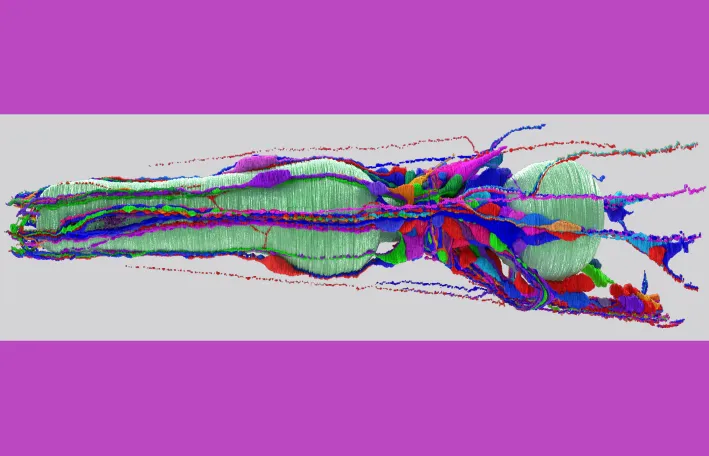
To enable comparisons between predator and prey nematodes, the Hobert lab team meticulously reconstructed a 3D wiring map of connections among all neurons in P. pacificus. This map, also known as a connectome, promises an understanding of the precise way in which information flows throughout a neural circuit to produce behavior. To track the paths neurons take throughout the body and the contacts they make along the way, Dr. Steven Cook and Christine Kalinski analyzed ultrathin tissue slices ~50 nm thick—about 2000 times thinner than a human hair—and imaged them at high resolution. These images were then lined up like the pages in a flipbook to reconstruct a 3D image. The real work could then begin–tracing each neuron across thousands of slices by hand. The researchers compared the finished connectome directly to the existing C. elegans connectome.
The researchers found the locations of neurons within the body as well as their basic features to be remarkably similar across species despite their vastly different behavioral outputs. “Even though we have known for a long time that nematode body plans are deeply conserved, I found the extent of conservation of neuronal cell typology over several hundred million years of evolution truly stunning,” said Dr. Oliver Hobert. “This being said, evolution and nature have found some nifty ways to modify circuit composition and architecture that we expect to explain these behavioral adaptations.”
One neuron shared across species that the researchers homed in on is the mechanical stimuli-sensing FLP cell. When touched on the head, C. elegans immediately retreat backwards to evade potential threats, an escape response driven by FLP touch detection. When touched, FLP signals to another neuron which prompts a backward movement. In P. pacificus, however, FLP is wired differently. Its axon is shorter and does not reach the backwards movement neuron. Instead, it signals to a neuron that promotes forward motion, meaning that rather than avoiding touch as C. elegans would, P. pacificus might move toward it. Cook and Kalinski posit that this rewiring, traced back to a single neuron, could promote P. pacificus’ distinctive predatory lifestyle. When P. pacificus worms track down and make physical contact with prey, they do not retreat; they keep moving forward to attack.
But not all connectome alterations drive behavioral change. While painful stimuli are perceived by the ASH neuron in both nematodes, this neuron travels along different sides of the worm’s central brain. As a result, it extends through different neuronal neighborhoods in the two species, presenting opportunities for species-specific synaptic connections. Yet, despite these structural differences, ASH elicits identical avoidance behavior in both species. Since behavioral differences cannot always be attributed to a single wiring change, broader network-level adaptations must be investigated to uncover how two superficially identical nematodes produce markedly different behaviors.
By comparing the brains of two nematode species whose evolution diverged millions of years ago, the team from the Hobert lab was able to identify which aspects of neuronal wiring have remained stable over millions of years and where new circuits have evolved. This impressive work offers insights into exactly how evolution alters the brain by the fine tweaking of neuronal synapses. Even small changes in connections between neurons or their positions with relation to neighbors can accumulate to produce species with disparate behavioral repertoires. This new research offers valuable clues into what makes a species tick—or in this case, hunt.